Researchers use extraembryonic tissue to produce entities that resemble human embryos

Massachusetts, US: In the "black box" of human development known as gastrulation, an embryo transforms from a hollow, spherical structure to a multilayered one. Human embryos are typically not cultured for longer than 14 days due to bioethical concerns, and gastrulation happens between 17 and 21 days after fertilisation.
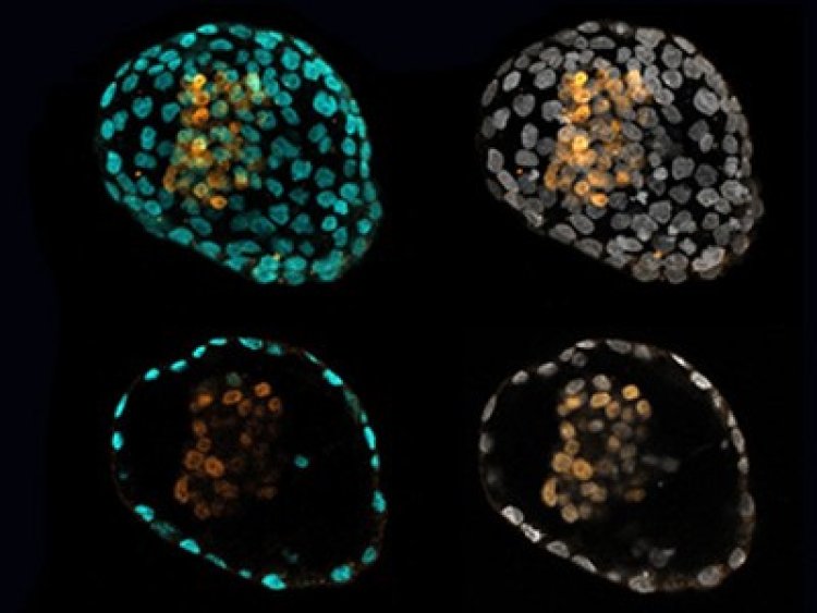
Furthermore, the extraembryonic parts that give rise to the yolk sac and placenta are not included in the current stem cell models that simulate gastrulation. In a study published in the journal Cell, researchers describe a novel method for creating "peri-gastruloids," an embryo-like structure that contains the yolk sac, one of the supporting tissues that was absent from earlier models.
“While non-integrated models of human gastrulation and early organogenesis have been developed from primed human pluripotent stem cells, these models lack the extraembryonic cells that play vital roles in embryo patterning and morphogenesis,” says senior author Jun Wu, a stem cell biologist at the University of Texas Southwestern Medical Center. “The presence of both embryonic and extraembryonic tissues enables researchers to examine the interactions between the epiblast, amnion, and yolk sac during gastrulation—an endeavour previously unattainable in humans.”
Instead of the more commonly used primed pluripotent stem cells, the researchers’ method used expanded pluripotent stem cells (EPSCs). These cells have previously been shown to differentiate into both embryonic and extraembryonic tissue in mice. By adding the proper growth factors to human EPSCs, they differentiated into these two types of tissues. The cells then self-organized into structures that resembled the human embryo, which the researchers refer to as “peri-gastruloids.”
Extraembryonic tissues release chemical signals that guide embryo development, which allows these peri-gastruloids to mimic several important processes that are considered part of this black-box period of development. Peri-gastruloids develop the amniotic cavity that embryos live inside, and the yolk sac cavities that provide the embryos with blood supply. In addition, peri-gastruloids show early signs of organogenesis, such as neurulation, which marks the very beginning of central nervous system development.
The research team reports that their method is efficient and reproducible. In what they consider a small-scale trial, they were able to generate hundreds of peri-gastruloids. “The power of this model stems from its ability to exploit the remarkable self-organizing capacity of human EPSCs with minimal external intervention” says Wu.
The team notes that peri-gastruloids are not viable because of the exclusion of trophoblasts that give rise to the placenta, which helps assuage the ethical concerns of this research. This project followed international stem cell research guidelines and was approved by UT Southwestern’s Stem Cell Oversight Committee.